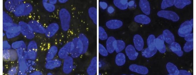
In oligodendrocytes with the APOE4 variant (left image), lipid droplets (yellow) accumulate. After treatment with a GSK3b inhibitor (right image), the lipid droplets disappear. Courtesy of the Picower Institute for Learning and Memory at the Massachusetts Institute of Technology (MIT), United States.
A molecular pathway has been identified through which a genetic risk factor that causes Alzheimer’s disease interferes with the formation of the fatty sheath that surrounds brain neurons. The study confirmed that inhibiting a protein involved in the process that disrupts sheath formation restores the sheath to normal, raising expectations that this could provide an important clue for the development of Alzheimer’s disease treatments.
A research team led by Professor Li-Huei Tsai of the Picower Institute for Learning and Memory at the Massachusetts Institute of Technology (MIT) in the United States identified the molecular pathway through which “APOE4 gene variants,” the strongest genetic risk factor for Alzheimer’s disease, damage the fatty sheath surrounding brain neurons. The team published its findings on the 15th (local time) in the international journal “Cell Stem Cell.”
Neurons in the brain transmit information by exchanging electrical signals. The long projections extending from neurons, known as “axons,” are wrapped in a fatty sheath called “myelin,” which acts like electrical insulation to prevent signals from leaking. Oligodendrocytes in the brain use lipids as raw material to produce myelin.
When myelin properly wraps axons, neural signals are transmitted quickly and accurately. In contrast, previous observations have shown that in the presence of APOE4 gene variants, myelin does not form correctly. If myelin does not form properly, neural signal transmission slows down or malfunctions.
Using CRISPR gene-editing technology, the research team systematically knocked out protein-coding genes one by one in cultured oligodendrocytes with APOE4 gene variants, in a search for the key switch that prevents myelin formation.
As a result, the team confirmed that suppression of the “Wnt signaling pathway” in oligodendrocytes interferes with myelin formation. The Wnt signaling pathway is an essential signaling system that regulates cell proliferation, differentiation, migration, and regeneration.
When the Wnt signaling pathway is blocked, oligodendrocytes can no longer utilize lipids to produce myelin. Consequently, unnecessary lipid droplets accumulate inside the oligodendrocytes.
The team also found that in the presence of APOE4 gene variants, levels of “beta-catenin,” a protein that promotes Wnt signaling, decrease, while “GSK3b,” a protein that suppresses Wnt signaling, is overexpressed. When a GSK3b-inhibiting drug was administered to a brain organoid (organoid) model, Wnt signaling was restored. The lipid droplets (intracellular fat storage depots) that had accumulated within the cells decreased, myelin formed normally, and axonal growth in neurons was observed to be promoted.
The drug’s effects were also confirmed in animal experiments. When an Alzheimer’s disease mouse model was treated with a GSK3b inhibitor, unnecessary lipid droplets in the hippocampus—a major brain region responsible for memory—decreased, and myelin again thickened.
The research team stated, “Through genetic screening, we identified the pathway of lipid metabolism defects triggered by Alzheimer’s disease risk factors,” adding, “The significance of this study lies not only in elucidating the molecular mechanism but also in presenting a new drug target for the development of Alzheimer’s disease treatments.”
ⓒ dongA.com. All rights reserved. Reproduction, redistribution, or use for AI training prohibited.
Popular News