Domestic researchers develop AI for automatic detection
Analysis of 1,060 three-dimensional brain MRI scans
Incidence reaches 66.7% in CADASIL patients
Volume of white matter degeneration about 1.7 times larger
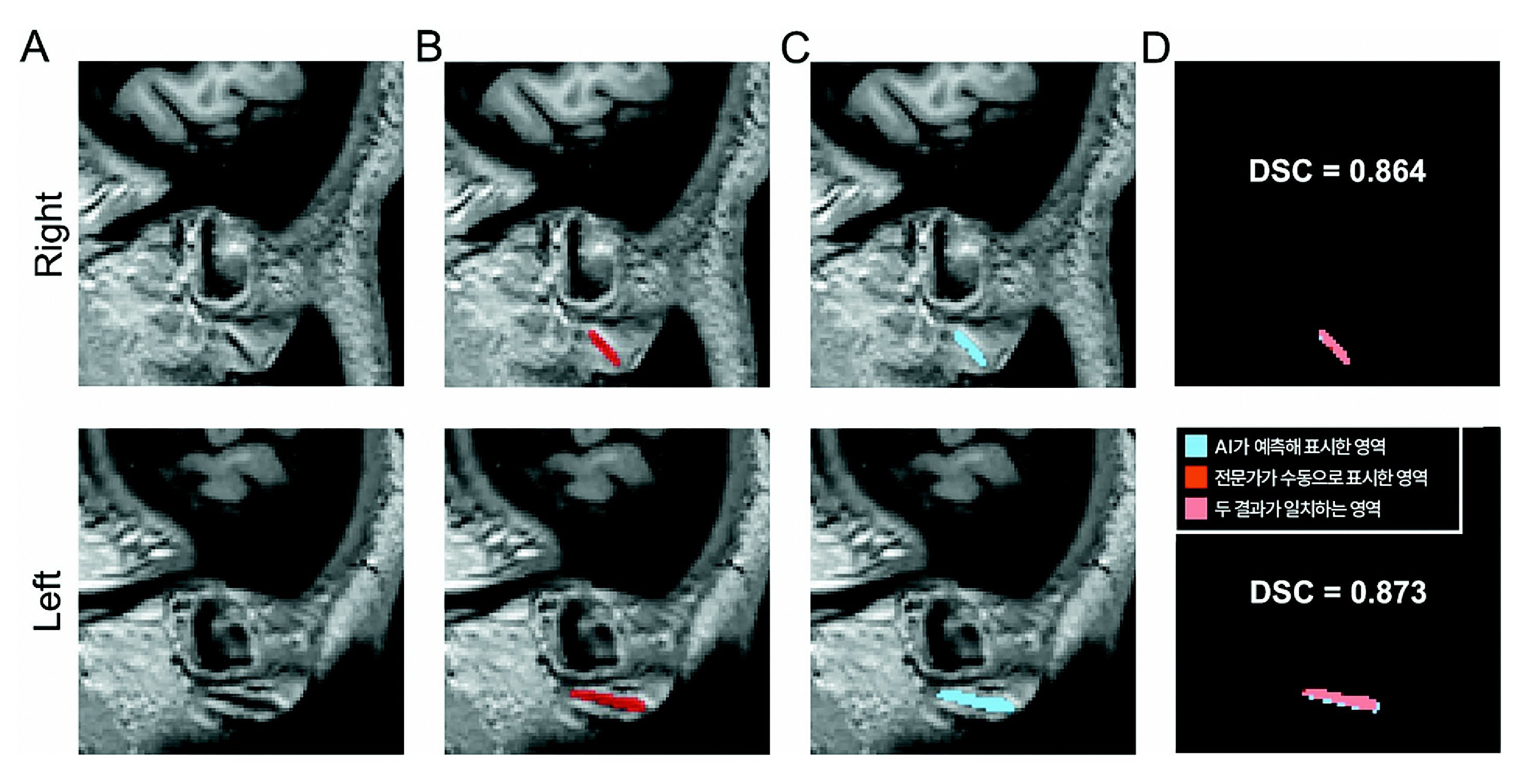
Based on the three-dimensional original image (A), wrinkles manually marked by an expert (B) and areas automatically marked by AI predictions (C).
Attention is required if a deeply indented diagonal crease is visible on the earlobe when looking in the mirror. It is easy to dismiss, but this fold, known as “Frank’s sign,” has recently attracted attention for its association with cardiocerebrovascular diseases. A comedian who suddenly collapsed during filming and was diagnosed with acute myocardial infarction recently drew public interest when his earlobe crease was noticed.
A Korean research team has presented scientific evidence that this may be a signal beyond a simple aging phenomenon by using artificial intelligence (AI) to objectively identify Frank’s sign and clarify its association with the severity of hereditary cerebral small vessel damage. Cerebral small vessels are very small (diameter 10–300 μm) blood vessels distributed throughout the brain that play a crucial role in supplying oxygen and nutrients to the brain.
Frank’s sign is a deep diagonal crease that appears at about a 45-degree angle on one or both earlobes. It was first reported in 1973 by American physician Sanders Frank, who observed that this fold frequently appeared in patients with angina pectoris. Subsequent reports indicated that it is relatively common in patients with cardiocerebrovascular diseases such as myocardial infarction, stroke, and vascular dementia. However, until now, only a correlation—“frequently observed in patients with vascular disease”—had been confirmed, and no clear causal relationship or mechanism of occurrence had been identified.
Another factor that complicated research was the subjectivity of assessment. Frank’s sign has been judged visually by researchers directly inspecting the ear or examining two-dimensional photographs, but in the absence of standardized definitions or criteria, the same patient could be evaluated differently depending on the assessor.
Frank’s sign objectified with AI beyond visual judgmentTo address these limitations, a team led by Professor Ki Woong Kim of the Department of Neuropsychiatry at Seoul National University Bundang Hospital developed the world’s first AI model that automatically detects Frank’s sign in three-dimensional brain magnetic resonance imaging (MRI). The team focused on the fact that the face, including both earlobes, is captured together during brain MRI scans and utilized three-dimensional facial images extracted from the MRI.
Based on 400 brain MRI scans collected at Seoul National University Bundang Hospital, experts manually annotated Frank’s sign, and these data were used to train the AI. The first validation was then conducted using 600 cases from the same hospital that were not used for training, and the second validation was performed using 460 multicenter cases from Chungnam National University Hospital, Kangwon National University Hospital, and Severance Hospital.
In the validation, the team compared the degree of overlap between the wrinkle regions marked by experts and the regions automatically segmented by AI. As a result, the DSC (Dice similarity coefficient), which represents the similarity between the two regions, was 0.734 and 0.714, respectively. In the field of medical imaging, values above 0.7 are considered highly accurate. The AUC (area under the curve), which reflects performance in distinguishing the presence or absence of Frank’s sign, also exceeded 0.9 in all cases, demonstrating that the model can operate reliably across various clinical settings.
Association revealed in hereditary cerebral small vessel diseaseThe research team went a step further and used the developed AI model to analyze whether Frank’s sign actually reflects the extent of vascular damage. The subjects were patients with CADASIL, a hereditary cerebral small vessel disease caused by a single-gene mutation. CADASIL is characterized by white matter hyperintensities (WMH), and as lesions accumulate, the risks of stroke and dementia increase.
When the team compared 81 CADASIL patients with genetically confirmed diagnoses to 54 age- and sex-matched healthy individuals, the prevalence of Frank’s sign in CADASIL patients was 66.7%, significantly higher than in healthy individuals (42.6%). Even after adjusting for age and other factors, CADASIL patients were 4.2 times more likely than healthy individuals to have Frank’s sign.
In addition, within the CADASIL patient group, those with Frank’s sign had a white matter hyperintensity volume approximately 1.7 times larger than those without it. When white matter hyperintensity volume was divided into lower, middle, and upper tertiles, the prevalence of Frank’s sign increased stepwise at 37.0%, 66.7%, and 74.1%. These findings suggest that Frank’s sign is associated with disease severity.
Professor Kim stated, “This study provides scientific evidence that the much-debated Frank’s sign is not merely a trace of aging but can objectively reflect the degree of hereditary cerebral small vessel damage,” adding, “Frank’s sign alone cannot be used to diagnose disease, but if there are vascular risk factors such as hypertension or diabetes, it is advisable to recognize an earlobe crease as an additional warning sign and seek consultation with a specialist.”
This study was conducted with support from the Ministry of Health and Welfare, the Korea Health Industry Development Institute, and the Dementia Research and Development Project Group and was published in the international journals ‘Scientific Reports’ and ‘Journal of Clinical Medicine’. Observers note that it is significant in that the debate around Frank’s sign is shifting from empirical observation to quantitative research.
ⓒ dongA.com. All rights reserved. Reproduction, redistribution, or use for AI training prohibited.
Popular News